Surgery on the distal bronchi. Treatment of bronchiectasis. Operations for bronchiectasis. Treatment of tracheal cancer
All materials on the site were prepared by specialists in the field of surgery, anatomy and specialized disciplines.
All recommendations are indicative in nature and are not applicable without consulting a doctor.
The need for lung surgery always causes reasonable fear in both the patient and his relatives. On the one hand, the intervention itself is quite traumatic and risky, on the other hand, operations on the respiratory organs are indicated for persons with serious pathology, which without treatment can lead to the death of the patient.
Surgical treatment of lung diseases places high demands on the general condition of the patient, since it is often accompanied by major surgical trauma and a long period of rehabilitation. Interventions of this kind should be taken seriously, paying due attention to both preoperative preparation and subsequent recovery.
The lungs are a paired organ located in the thoracic (pleural) cavities. Life without them is impossible, because the main function of the respiratory system is to deliver oxygen to all tissues of the human body and remove carbon dioxide. At the same time, having lost part or even the whole lung, the body can successfully adapt to new conditions, and the remaining part of the pulmonary parenchyma is able to take on the function of the lost tissue.
The type of lung surgery depends on the nature of the disease and its prevalence. If possible, surgeons preserve the maximum volume of respiratory parenchyma, unless this contradicts the principles of radical treatment. In recent years, modern minimally invasive techniques have been successfully used to remove lung fragments through small incisions, which contributes to a faster recovery and a shorter recovery period.
When is lung surgery necessary?
Lung operations are performed if there is a serious reason for this. Indications include:
The most common causes of lung surgery are tumors and some forms of tuberculosis. For lung cancer, surgery includes not only the removal of a part or an entire organ, but also excision of the lymphatic drainage pathways - the intrathoracic lymph nodes. In case of extensive tumors, resection of the ribs and areas of the pericardium may be required.

types of operations for surgical treatment of lung cancer
The types of lung interventions depend on the amount of tissue removed. Thus, a pulmonectomy is possible - removal of an entire organ, or resection - excision of a fragment of the lung (lobe, segment). With the widespread nature of the lesion, massive cancer, disseminated forms of tuberculosis, it is impossible to rid the patient of the pathology by removing only a fragment of the organ, therefore radical treatment is indicated - pneumonectomy. If the disease is limited to a lobe or segment of the lung, then it is enough to excise only them.
Traditional open surgeries are performed in cases where the surgeon is forced to remove a large volume of an organ. Recently, they are giving way to minimally invasive interventions, which make it possible to excise the affected tissue through small incisions - thoracoscopy. Among modern minimally invasive methods of surgical treatment, the use of laser, electric knife, and freezing are gaining popularity.
Features of operations
When performing interventions on the lung, accesses are used that provide the shortest path to the pathological focus:
- Anterolateral;
- Side;
- Posterolateral.
Anterolateral approach means an arcuate incision between the 3rd and fourth ribs, starting slightly lateral from the parasternal line, extending to the posterior axilla. Posterolateral lead from the middle of the third or fourth thoracic vertebrae, along the paravertebral line to the angle of the scapula, then along the sixth rib to the anterior axillary line. Side cut is carried out when the patient lies on the healthy side, from the midclavicular line to the paravertebral line, at the level of the fifth-sixth rib.

Sometimes, in order to reach the pathological focus, sections of the ribs have to be removed. Today it has become possible to excise not only a segment, but also an entire lobe thoracoscopically, when the surgeon makes three small incisions of about 2 cm and one up to 10 cm, through which instruments are inserted into the pleural cavity.
Pulmonectomy
Pulmonectomy is an operation to remove the lung, which is used in cases of damage to all its lobes in common forms of tuberculosis, cancer, and purulent processes. This is the most significant operation in terms of volume, because the patient loses an entire organ at once.
 The right lung is removed from the anterolateral or posterior approach. Once in the chest cavity, the surgeon first ties up the elements of the lung root individually: first the artery, then the vein, and the bronchus is the last to be ligated. It is important that the bronchial stump is not too long, because this creates a risk of stagnation of contents, infection and suppuration, which can cause failure of the sutures and inflammation in the pleural cavity. The bronchus is stitched with silk or sutures are applied using a special device - a bronchial stitcher. After ligating the elements of the lung root, the affected organ is removed from the chest cavity.
When the bronchial stump is sutured, it is necessary to check the tightness of the sutures, which is achieved by pumping air into the lungs. If everything is in order, then the area of the vascular bundle is covered with pleura, and the pleural cavity is sutured, leaving drainage in it.
The left lung is usually removed through an anterolateral approach. The left main bronchus is longer than the right, so the doctor must be careful that its stump does not turn out to be long. The vessels and bronchus are treated in the same way as on the right side.
Pulmonectomy (pneumonectomy) is performed not only on adults, but also on children, but age does not play a decisive role in the choice of surgical technique, and the type of operation is determined by the disease (bronchiectasis, polycystic lung disease, atelectasis). In case of severe pathology of the respiratory system, requiring surgical correction, expectant management is not always justified, since many processes can disrupt the growth and development of a child if not treated in a timely manner.
Lung removal is performed under general anesthesia. It is necessary to administer muscle relaxants and tracheal intubation for ventilation of the organ parenchyma. In the absence of an obvious inflammatory process, drainages may not be left, and the need for them arises when pleurisy or other effusion appears in the chest cavity.
Lobectomy
A lobectomy is the removal of one lobe of the lung, and if two lobes are removed at once, the operation will be called a bilobectomy. This is the most common type of lung surgery. Indications for lobectomy are tumors limited to a lobe, cysts, some forms of tuberculosis, and isolated bronchiectasis. Lobectomy is also performed in cases of oncopathology, when the tumor is local in nature and does not spread to surrounding tissues.

lobectomy
The right lung includes three lobes, the left - two. The upper and middle lobes of the right and the upper lobe of the left are removed from the anterolateral approach, the lower lobe of the lung is removed from the posterolateral one.
After opening the chest cavity, the surgeon finds the vessels and bronchus, ligating them separately in the most minimally traumatic manner. First, the vessels are treated, then the bronchus, which is sutured with a thread or bronchial stitch. After these manipulations, the bronchus is covered with pleura, and the surgeon removes a lobe of the lung.
After a lobectomy, it is important to straighten the remaining lobes during surgery. To do this, oxygen is pumped into the lungs under high pressure. After the operation, the patient will have to independently straighten the lung parenchyma by performing special exercises.
After lobectomy, drains are left in the pleural cavity. During upper lobectomy, they are installed through the third and eighth intercostal space, and when removing the lower lobes, one drain inserted into the eighth intercostal space is sufficient.
Segmentectomy
A segmentectomy is an operation to remove part of the lung, called a segment.. Each lobe of the organ consists of several segments that have their own artery, vein and segmental bronchus. It is an independent pulmonary unit that can be excised safely for the rest of the organ. To remove such a fragment, use any of the approaches that provide the shortest possible path to the affected area of the lung tissue.
Indications for segmentectomy include small lung tumors that do not extend beyond the segment, lung cysts, small segmental abscesses and tuberculous cavities.
After dissecting the chest wall, the surgeon isolates and ligates the segmental artery, vein, and lastly the segmental bronchus. Isolation of a segment from the surrounding tissue should be done from the center to the periphery. At the end of the operation, drainage is installed in the pleural cavity according to the affected area, and the lung is inflated with air. If a large number of gas bubbles are released, the lung tissue is sutured. X-ray control is required before closing the surgical wound.
Pneumolysis and pneumotomy
Some operations on the lungs are aimed at eliminating pathological changes, but are not accompanied by the removal of its parts. These include pneumolysis and pneumotomy.

Pneumolysis is an operation to cut adhesions that prevent the lung from expanding and filling with air. A strong adhesive process accompanies tumors, tuberculosis, suppurative processes in the pleural cavities, fibrinous pleurisy in kidney pathology, extrapulmonary neoplasms. Most often, this type of operation is performed for tuberculosis, when abundant dense adhesions are formed, but the size of the cavity should not exceed 3 cm, that is, the disease should be limited in nature. Otherwise, a more radical intervention may be required - lobectomy, segmentectomy.
Dissection of adhesions is carried out extrapleurally, intrapleurally or extraperiosteally. At extrapleural In pneumolysis, the surgeon peels off the parietal pleural layer (outer) and introduces air or petroleum jelly into the chest cavity to prevent the lung from inflating and the formation of new adhesions. Intrapleural dissection of adhesions is carried out by penetration under the parietal pleura. Extraperiosteal the method is traumatic and has not been widely used. It involves peeling off the muscle flap from the ribs and introducing polymer beads into the resulting space.
The adhesions are cut using a hot loop. The instruments are inserted into the part of the chest cavity where there are no adhesions (under X-ray control). To gain access to the serous membrane, the surgeon resects sections of the ribs (the fourth for upper lobe lesions, the eighth for lower lobe lesions), peels off the pleura and sutures the soft tissue. The entire treatment process takes up to one and a half to two months.

lung abscess
Pneumotomy is another type of palliative surgery, which is indicated for patients with focal purulent processes - abscesses. An abscess is a cavity filled with pus, which can be evacuated by opening the chest wall.
Pneumotomy is also indicated for patients with tuberculosis, tumors and other processes that require radical treatment, but which is impossible due to their serious condition. Pneumotomy in this case is intended to make the patient feel better, but will not help completely eliminate the pathology.
Before performing a pneumotomy, the surgeon must perform a thoracoscopy to find the shortest path to the pathological focus. Then the rib fragments are resected. When access to the pleural cavity is obtained and provided that there are no dense adhesions in it, the latter is tamponed (the first stage of the operation). After about a week, the lung is dissected, and the edges of the abscess are fixed to the parietal pleura, which ensures the best outflow of pathological contents. The abscess is treated with antiseptics, leaving tampons soaked in a disinfectant in it. If there are dense adhesions in the pleural cavity, then pneumotomy is performed in one stage.
Before and after surgery
Surgeries on the lungs are traumatic, and the condition of patients with pulmonary pathology is often severe, so proper preparation for the upcoming treatment is very important. In addition to standard procedures, including a general blood and urine test, biochemical blood test, coagulogram, and lung x-ray, CT, MRI, fluoroscopy, and ultrasound examination of the chest organs may be required.

In case of purulent processes, tuberculosis or tumors, by the time of the operation the patient is already taking antibiotics, anti-tuberculosis drugs, cytostatics, etc. An important point in preparing for lung surgery is breathing exercises. In no case should it be neglected, since it not only promotes the evacuation of contents from the lungs even before the intervention, but is also aimed at straightening the lungs and restoring respiratory function after treatment.
In the preoperative period, a physical therapy methodologist helps you perform exercises. A patient with abscesses, cavities, or bronchiectasis should turn and bend the torso while simultaneously raising the arm. When the sputum reaches the bronchus and causes a cough reflex, the patient leans forward and down, facilitating its removal with a cough. Weakened and bedridden patients can perform exercises while lying in bed, with the head end of the bed lowered slightly.
 Postoperative rehabilitation takes on average about two weeks, but can last longer, depending on the pathology. It includes treatment of the postoperative wound, changing bandages, tampons for pneumotomy, etc., adherence to the regimen and exercise therapy.
The consequences of the treatment may include respiratory failure, secondary purulent processes, bleeding, suture failure and pleural empyema. To prevent them, antibiotics and painkillers are prescribed, and discharge from the wound is monitored. Breathing exercises are required, which the patient will continue to perform at home. The exercises are performed with the help of an instructor, and should be started within a couple of hours from the moment you emerge from anesthesia.
Life expectancy after surgical treatment of lung diseases depends on the type of intervention and the nature of the pathology. Thus, when single cysts, small tuberculosis lesions, and benign tumors are removed, patients live as long as other people. In the case of cancer, severe purulent process, lung gangrene, death can occur from septic complications, bleeding, respiratory and heart failure at any time after the intervention, if it did not contribute to achieving a stable condition.
If the operation is successfully performed and there are no complications or progression of the disease, the prognosis is generally good. Of course, the patient will need to monitor his respiratory system, smoking is out of the question, breathing exercises will be needed, but with the right approach, healthy lobes of the lungs will provide the body with the necessary oxygen.
Disability after lung surgery reaches 50% or more and is indicated for patients after pneumonectomy, in some cases after lobectomy, when the ability to work is impaired. The group is assigned in accordance with the patient's condition and is periodically reviewed. After a long period of rehabilitation, the majority of those operated on regain both health and ability to work. If the patient has recovered and is ready to return to work, then the disability can be lifted.
Lung operations are usually performed free of charge, because this is required by the severity of the pathology, and not by the patient’s desire. Treatment is available in thoracic surgery departments, and many operations are performed under the compulsory medical insurance system. However, the patient can undergo paid treatment in both public and private clinics, paying for both the operation itself and comfortable conditions in the hospital. The cost varies, but it cannot be low, because lung surgery is complex and requires the participation of highly qualified specialists. On average, pneumonectomy costs about 45-50 thousand, and for excision of mediastinal lymph nodes - up to 200-300 thousand rubles. Removing a lobe or segment will cost from 20 thousand rubles in a public hospital and up to 100 thousand in a private clinic.
Or rather, its tactics directly depend on the degree of development of the oncological process, its form, the presence of metastasis to nearby organs and tissues, and the effectiveness of histological tests. In 80% of cancer patients, radical intervention has proven its effectiveness; in more than 30% of patients with lymph nodes affected, surgery prolongs life.
How is surgery performed for bronchial cancer?
Depending on the indications, treatment is carried out as follows: Removal of part of the lung: lobectomy - resection of one lobe of the organ, bilobectomy - excision of two lobes of the lung; Complete resection of the lung: pneumonectomy, pneumonectomy with mediastinal lymphadenectomy, excision of a segment of the trachea in the area of its division into the main branches with restoration of the integrity of the tracheobronchial tree, pneumonectomy with circular resection of the superior vena cava/thoracic aorta. In some cases, when a cancerous process is detected in the early stages, a circular or fenestrated removal of the organ is performed. To combat the oncological process, surgical or combined methods are used. Combined includes the use of gamma therapy. The cost of surgery for bronchial cancer using gamma therapy depends on the course of the pathological process, its stages. Cancer, as a rule, metastasizes to nearby lymph nodes in a short time, so any intervention involves excision of those lymph nodes that receive lymph from the affected organ. This approach to treatment minimizes the risk of possible progression of the pathology and relapses, which significantly increases the life expectancy of patients. The intervention begins with opening the chest (thoracotomy), followed by fixation of the vessels, cauterization and suturing. After this, the surgeon removes the required area of the lung, or performs a complete resection of the organ, fatty tissue and lymph nodes, and then creates a stump. The cavity part in which the lung was located undergoes restoration and reduction. At the end of the procedure, drainage is installed and sutures are applied. The patient is transferred to the intensive care unit, where artificial ventilation (ALV) will be used until the person emerges from anesthesia.
How much does surgery to remove bronchial cancer cost?
In various Moscow clinics, the cost of surgery to remove bronchial cancer is determined on an individual basis. The cost includes diagnostic measures, the extent of surgical intervention, and the duration of the postoperative period. You can make an appointment with a qualified specialist or fill out an application for surgical treatment on our website.
Postoperative period for bronchial cancer
Rehabilitation after surgery for bronchial cancer involves careful monitoring of the patient’s condition, blood pressure control, respiratory function. In the first few days, active aspiration of the pleural cavity is carried out using drainage and suction. The patient is prescribed a course of antibiotic therapy and other medications, depending on the outcome of the intervention and possible adverse consequences. Gentle dietary nutrition is necessary in the first two to three days. The patient is taught special breathing exercises, which improve blood circulation and prevent congestion in the lungs and bronchi. Consequences after surgery for bronchial cancer can manifest themselves in the form of relapses of oncological processes in the remaining part of the organ, but only if there is infiltration of the bronchial wall behind the visible outlines of the tumor formation.
The lungs are a unique paired organ that provides our entire body with vital oxygen and removes carbon dioxide from it. And although they are often susceptible to various diseases, their compensatory capabilities are great, and the tissue is very plastic. This allows you to perform various surgical interventions, up to the complete removal of one lung.
The lungs have a unique structure. Due to the presence of half a billion alveoli (respiratory vesicles), an organ weighing only 1 kilogram has a total respiratory surface area of 100-150 square meters, which can be compared to the area of a tennis court. During the day, this surface passes and “conditions” more than 10,000 liters of air. The length of the respiratory tract, that is, the branched bronchial tree, is about 3,000 kilometers. Moreover, the amount of blood passing through the lungs per day is 6-7 tons!
The lungs, like the heart, work “automatically”; this process is regulated by a complex neuro-humoral mechanism. And although we cannot temporarily stop cardiac activity through an effort of will, like breathing, its stop is also short-lived, because the automatic mechanisms of inhalation are activated, regardless of our will.
In the alveoli of the lungs there is a so-called air reserve of about 200 ml. In emergency situations, it is included in the breathing process, and normally it is periodically updated in the form of deep sighs and yawns.
Lung diseases that require surgery
Unfortunately, not all lung diseases can be cured with medications and procedures. Many of them can only be treated with surgery:

All these operations are performed in specialized departments of thoracic (chest) surgery by highly qualified specialists.
Advice: Often the most dangerous lung diseases, including cancer, can begin with a seemingly harmless cough. It cannot be ignored; you must consult a doctor and undergo an examination.
Types of lung operations
All interventions on the lungs can be divided into 2 groups according to volume: pneumonectomy or pneumonectomy (complete removal of the lung) and resection (removal of part of the lung). Complete removal is performed in cases of malignant tumors, as well as in cases of multiple pathological foci in all lobes of the organ.
Lung resection can be of various volumes:
- atypical or marginal - removal of a limited area on the periphery;
- segmentectomy – removal of a segment with the corresponding segmental bronchus;
- lobectomy – removal of one lobe;
- bilobectomy – removal of 2 lobes;
- reduction - a decrease in lung volume when they are affected by emphysema (non-functioning air cavities in the organ tissue).

According to technology, all interventions are of 2 types: thoracotomy or traditional - with a wide opening of the chest, and thoracoscopic - minimally invasive, performed using endovideo technology.
Surgical procedures also include thoracentesis. It can be performed with a needle, as well as, or by making a small incision with the insertion of a drainage tube to drain fluid (pus, blood) to administer medications.
Finally, the most difficult operation is a lung transplant, which today is quite often performed both abroad and in large domestic clinics in cases where both lungs fail.
Modern technologies of pulmonary surgery
Thanks to the emergence of unique innovative surgical technologies, many lung surgeries are performed today using a minimally invasive method through several small incisions on the skin no more than 3 cm long. The technology is similar through small incisions with a video camera. In principle, the technology of thoracoscopic operations is the same as for laparoscopic interventions on the abdominal organs (, intestines, and others).
Such operations are much less traumatic, short in duration, do not require long hospitalization, and the rehabilitation period is much shorter.
Among the innovative methods, laser surgery is also used to remove tumors, including cancer, as well as radiosurgery, cryodestruction (freezing). All these technologies are performed minimally invasively - through percutaneous puncture, bronchoscopy or thoracoscopy.
Advice: If you are going to have lung surgery, you must stop smoking in advance and do breathing exercises so that your lungs can cleanse themselves. Smokers are much more likely to experience post-operative complications.
Postoperative rehabilitation

Removal of a lung or part of it inevitably leads to disruption of respiratory function and oxygen metabolism of the entire body. The main task of the recovery period is to “breathe” the remaining volume of the lungs, taking into account their compensatory capabilities and ensure normal gas exchange.
In the early postoperative period, special methods are used in the hospital - hardware, medications, exercise therapy, inhalations - according to individual schemes for each patient. After discharge, these tasks are assigned to the patient himself. The main activities are:
- general hygienic gymnastics;
- special breathing exercises;
- visiting physiotherapeutic procedures, inhalations;
- maintaining a diet with sufficient protein and vitamins;
- regular walks in the fresh air.
Lung operations today are performed using new, gentle technologies, and in combination with professional postoperative rehabilitation, they give good results in restoring respiratory function.
Video
Attention! The information on the site is presented by specialists, but is for informational purposes only and cannot be used for independent treatment. Be sure to consult your doctor!
Choice of treatment method depends on the prevalence of bronchiectasis, age and general condition of patients. With strictly localized, including bilateral, bronchiectasis, practical recovery is possible only after surgical treatment. Scattered, polysegmental unilateral and bilateral bronchiectasis are subject to therapeutic treatment. Indications for surgery should be especially careful in children under 7 years of age, since they have a tendency to generalize purulent bronchitis and it is difficult to determine the boundary between functional and organic changes in the bronchi.
Must be discreet attitude towards surgical treatment in patients over 45-50 years old. Along with bronchiectasis, even strictly localized ones, diffuse pneumosclerosis occurs in such patients as a result of a long suppurative process. On the one hand, surgery in such patients poses a certain risk; on the other hand, functional results after resection of part of the lung are often unsatisfactory.
Difficult to resolve the issue of testimony for surgery for bilateral bronchiectasis. It is fundamentally important to emphasize that the affected parts of the lung do not participate in gas exchange. Their resection itself does not cause significant functional impairment, which makes it possible to remove up to 13 segments of the lungs. However, an indispensable condition is to leave at least 3 segments in each lung. The operation is contraindicated in two cases: with total damage to one lung and bronchiectasis in the other lung and with a diffuse type of bronchial damage without clear localization. Experience shows that with bilateral bronchiectasis, the best long-term results are observed in patients operated on in childhood and adolescence. At the age of over 25-30 years, the indications for surgical treatment become narrower. The operation is contraindicated in the presence of severe pulmonary and heart failure, significant pneumosclerosis in segments free from bronchiectasis, severe purulent intoxication and amploplosis of internal organs.
Correct definition of indications and contraindications for surgical treatment of bronchiectasis largely depends on the results of previous complex therapy. The main attention is paid to the sanitation of the bronchial tree (bronchoscopy, bronchial catheterization under local anesthesia, aerosol inhalation, postural drainage). Along with this, restorative and symptomatic treatment is carried out. This tactic often gives a good therapeutic effect, and patients who seemed inoperable tolerate surgery well. In the preoperative period, sanitation of the tracheobronchial tree and restorative treatment are also carried out. It is important to achieve maximum suppression of inflammation in the bronchi and reduce the amount of sputum to 50-70 ml per day. If sanitation of the bronchi does not give the desired result within 2-3 weeks, it should be stopped, since increasing the duration of treatment may adversely affect the general condition of the patients. The safety of the operation largely depends on the method of anesthesia. Anesthetic tactics depend on the initial disturbances in gas exchange and the amount of sputum discharge.
Intubation anesthesia with muscle relaxants meets all the requirements of modern anesthesia. It ensures adequate gas exchange and prevents sputum from entering healthy parts of the lungs. At the same time, separate intubation of the bronchi allows the most reliable prevention of sputum aspiration.
Volume of lung resection determined on the basis of bronchography data. Operative diagnosis is complex and unreliable, therefore, in doubtful cases, it is preferable to remove the segment in which bronchial deformation is detected on the bronchogram even without obvious bronchiectasis. Leaving the diseased area of the lung causes a relapse of the disease. A typical operation for bronchiectasis is lobectomy, which is often combined with resection of the lingular segments or the middle lobe. Less commonly (mainly in childhood and adolescence) segmentectomy is performed. Even less often (in about 4-5% of cases) the entire lung is removed. For bilateral bronchiectasis, no more than 6 segments of each lung are resected. In rare cases, when conservative treatment does not give a lasting positive result, a large amount of purulent sputum and intoxication persist, palliative resection of the most affected area of the lung can be undertaken. Operations such as pneumotomy or thoracoplasty are not currently performed.
In the postoperative period the main attention is paid to bronchial patency. In all patients with bronchiectasis, the bronchial mucosa of the resected lung is prone to hypersecretion, therefore, already in the first hours after surgery, the bronchi should be actively emptied of contents. Patients are forced to cough, inhaled bronchodilators are prescribed, and the chest is massaged. If the patient is unable to independently cough up sputum, they resort to transnasal catheterization of the trachea and bronchi under fluoroscopy control. Obstruction of the bronchi with sputum for several hours inevitably leads to atelectasis of the operated lung, which may require bronchoscopy and bronchial lavage.
Of great importance adequate drainage of the pleural cavity. After partial resection of the lung, as a rule, air leaks through the resection surface for some time, which can lead to collapse of the pulmonary parenchyma, therefore, during the first day, drains are connected to active aspiration. After pneumonectomy, the pleural cavity is drained according to Bulau to control possible intrapleural bleeding. Anti-inflammatory preventive treatment is not necessary, since in the preoperative period and at the previous stages of treatment, patients receive large doses of various medications. Antibiotics are prescribed in case of infectious complications.
Prognosis for surgical treatment of bronchiectasis favorable. With unilateral, strictly localized (especially atelectatic forms) broichectasis, practical recovery is achieved in 75-85% of patients. Satisfactory results, i.e. reduction of cough and sputum, reduction of relapses of inflammation, are observed in 8-10% of those operated on. Unsatisfactory outcomes account for 3-5% of operations. It is important to emphasize that surgical treatment of broichiectasis should be considered as a stage of complex therapy. The best results are obtained when both therapists and surgeons participate equally in treatment. After the operation, clinical observation and periodic sanatorium-resort treatment are necessary.
Surgical treatment of pulmonary tuberculosis
In the complex of modern methods of treating patients with pulmonary tuberculosis, timely surgical interventions are important, and sometimes decisive. Over the past two decades, due to the successes of antibacterial therapy, anesthesiology and thoracic surgery, the possibility of using and the range of surgical interventions for pulmonary tuberculosis has expanded significantly, and the therapeutic effectiveness of operations has increased.
Numerous surgical interventions used for pulmonary tuberculosis can be classified as follows.
- Operations correcting artificial pneumothorax: a) thoracoscopy and thoracocaustics, b) open intersection of adhesions.
- Collapse-therapeutic operations: a) extrapleural pneumolysis with pneumothorax, filling and oleothorax, b) thoracoplasty.
- Lung resection. A. Cavern operations: a) cavity drainage, b) cavernotomy.
- Operations on the bronchi: a) ligation of the bronchus, suturing and dissection of the bronchus, b) resection and plastic surgery of the bronchus.
- Operations on the pulmonary vessels: a) ligation of the pulmonary veins, b) ligation of the pulmonary arteries.
- Operations on the nervous system: a) operations on the phrenic nerve, b) operations on the intercostal nerves.
- Decortication of the lung and pleurectomy.
- Removal of caseous lymph nodes.
Of these operations, some are performed frequently (lung resection, thoracoplasty), others are performed extremely rarely (surgeries on nerves and blood vessels). For all surgical interventions for pulmonary tuberculosis, complex treatment is carried out in the preoperative and postoperative periods in the form of a hygienic and dietary regimen and the use of antibacterial drugs. When appropriate, stimulating, desensitizing and hormonal therapy is also carried out. Let's consider the most important surgical interventions for pulmonary tuberculosis.
Thoracoscopy and thoracocaustics
One of the important methods of collapse therapy for pulmonary tuberculosis is artificial pneumothorax, which is often ineffective due to the presence of various intrapleural adhesions that prevent concentric collapse of the lung. In the absence of a positive clinical effect, treatment with artificial pneumothorax is inappropriate: adhesions contribute to the spread and exacerbation of the tuberculosis process, making the bloom of an anatomically defective pneumothorax dangerous.
In 1910-1913 Swedish phthisiatrician Jacobeus designed and used a special instrument with an optical system and a small light bulb at the end - a thoracoscope - to examine the pleural cavity. Soon a galvanocauter was added to the thoracoscope. With the help of these instruments, in the presence of a sufficient intrapleural gas bubble, it was possible to examine the pleural cavity in detail and burn the pleural cords under thoracoscopic control. This operation of closed burning of pleural adhesions is called thoracocaustics.
In the USSR, M. P. Umansky successfully performed thoracocaustics (1929); K. D. Esipov and especially the founder of Soviet phthisiosurgery N. G. Stoiko did a lot to improve and promote the burning of adhesions. In a short period of time, thoracocaustics was mastered by hundreds of surgeons and phthisiatricians in our country, becoming a method “without which artificial pneumothorax loses half of its value” (N. G. Stoiko).
At first, thoracocaustic surgery was accompanied by a significant number of complications, the main ones being bleeding and damage to the lung tissue. Over time, pleural adhesions were studied in detail, indications for surgery were clarified, instrumentation was improved, and the surgical technique of intervention was improved.
A comparison of clinical, radiological and thoracoscopic data shows that only thoracoscopy can provide a reliable idea of the presence, quantity, nature and operability of adhesions. The number of adhesions detected during thoracoscopy is always greater than that determined by x-ray examination. Therefore, thoracoscopy should be considered fundamentally indicated in every case of pneumothorax, taking into account the negative role of adhesions (N. G. Stoyko, A. N. Rozanov, A. A. Glasson, etc.)
Sometimes indications for thoracoscopy may be urgent. This applies to cases of stretching by cords of a thin-walled, subpleurally located cavity, with pulmonary hemorrhages that increase after inflation, spontaneous pneumothorax, if there is reason to assume that the cause of the rupture of the lung tissue was the fusion that fixed it.
The widespread use of antibacterial drugs has significantly reduced the risk of infection outbreaks after thoracoscopy and thoracocaustics. However, thoracoscopy should not be done in case of acute pulmonary process and acute pneumatic pleurisy. The presence of purulent exudate or tubercular lesions of the pleura is a contraindication to burning of adhesions. The most favorable time for the operation is 3-5 weeks after the application of an artificial pneumothorax.
Before performing thoracoscopy and thoracocaustics, the gas bubble in the pleural cavity must be sufficient in volume for the instruments to work freely: it must occupy at least a third of the pulmonary field. Points for insertion of instruments are marked before surgery, performing fluoroscopy in different positions of the patient. The pressure in the pleural cavity should be brought to atmospheric or close to it.

It is convenient to perform thoracoscopy and thoracocaustics in a darkened operating room. In this case, local anesthesia is usually used. After inserting a thoracoscope through the intercostal space, the condition of the pleura and lung is examined, and existing adhesions are examined.
The ability to navigate the thoracoscopic picture, understand the anatomical structure of the adhesions and determine the possibility of their burning is the most difficult part of the operation. If, after examining the pleural cavity, a decision is made to burn out the adhesions, a second instrument is introduced - a galvanocauter. The cautery loop is hidden in a special metal case placed on it. After bringing the cautery to the fusion, the loop is pulled out, the current is turned on, and the adhesion is burned out with a heated loop. The effect of the intervention can be noted not only during thoracoscopy, but also radiographically (Fig. 90 and 91).
When burning adhesions, special care is taken with respect to large vessels of the thoracic cavity (subclavian artery, aorta, etc.) and lung tissue involved in the adhesion. The rule is to burn the fusion after precise topographic-anatomical orientation, and as close to the chest wall as possible. Currently, thoracocautery is used much less frequently than in the 30-40s, since the indications for artificial pneumothorax have been narrowed.
Extrapleural pneumolysis with pneumothorax, filling and oleothorax
Extrapleural pneumolysis refers to the detachment of the parietal pleura and lung from the fascia lining the inside of the chest cavity.
In 1910, Tuffier and Martin proposed injecting air or nitrogen into the cavity formed after such lung detachment in tuberculosis and abscess. Insufflations were unsuccessful, after which Tuffier began filling the cavity with a fat filling, and Ver with paraffin. Later, other filling materials were tried (pieces of ribs, preserved cartilage, celluloid balls, methyl methacrylate balls, etc.). N. G. Stoiko paid a lot of attention to the method of extrapleural pneumolysis followed by filling with paraffin.
Due to frequent complications, extrapleural pneumolysis with filling is now rarely used. The filling is usually inserted extramusculo-periosteally, i.e. between the ribs on one side, exfoliated to the lung, and the costal periosteum and intercostal muscles on the other. Extrapleural pneumolysis with subsequent maintenance of an air bubble between the chest wall and the parietal pleura - extrapleural pneumothorax - has become more widespread.
To create extrapleural pneumothorax, more extensive pneumolysis is required than for filling. The operation is usually performed from the posterior or axillary approach after resection of a small portion of the rib. The lung is peeled off in front to the third rib, behind - to the VI-VII rib, laterally - to the IV rib and medially - to the root. After stopping minor bleeding, the chest cavity is hermetically sutured. As a rule, patients tolerate this relatively low-traumatic operation well.

Postoperative management of extrapleural pneumothorax is quite difficult, especially at first, and requires some experience. After the intervention, the lung tends to expand, and bloody fluid accumulates in the artificially formed cavity. In order to avoid this, during systematic X-ray monitoring, punctures are made to extract fluid and, if indicated, additional air is injected into the extrapleural cavity. As the gas bubble forms, the bottom of the extrapleural cavity gradually acquires a concave shape (Fig. 92). When extravasation no longer accumulates and there is a sufficient volume of air bubble, management of extrapleural pneumothorax becomes quite simple. By this time, the patient can be transferred for further treatment to a phthisiatrician and be under outpatient observation.
If the extrapleural cavity tends to shrink or it is impossible to maintain a gas bubble for some other reason, you can replace the air with oil, that is, switch to oleothorax (Fig. 93). The most suitable in these cases is Vaseline oil (300-400 ml), which after sterilization is introduced in several stages, removing the appropriate amount of air or liquid from the cavity. Vaseline oil dissolves very slowly, so it is usually not necessary to add it for several months. Transfer to oleothorax and the addition of oil are carried out under stationary conditions: the introduction of oil under high pressure is dangerous, as it can cause perforation of the lung and fat embolism.
The duration of treatment for extrapleural pneumothorax and oleothorax depends on the nature of the process for which the operation was performed and the condition of the cavity. With a smooth course of extrapleural pneumothorax imposed due to a fresh process, the gas bubble should be maintained for 1.5-2 years. Oleothorax in such cases should not last more than 3 years (T. N. Khrushcheva). After this period, it is necessary to periodically extract the oil in separate portions.
During the treatment of extrapleural pneumothorax, complications may occur in the form of the appearance of exudates in the cavity, specific and nonspecific suppurations, and the formation of internal bronchial fistulas. A fairly rare but dangerous complication is air embolism. With oleothorax, oil can penetrate the soft tissue of the chest wall or break into the bronchus. The latter is manifested by a cough and the release of petroleum jelly with sputum. To avoid aspiration of oil and the development of pneumonia in such cases, it is necessary to inculate the cavity and suck out the oil. Further treatment consists of resection and decortication of the lung or opening of the cavity, its sanitation and subsequent thoracoplasty.
In case of a stable immunobiological state of the body and obliteration of the pleural cavity, the indication for extrapleural pneumothorax is unilateral upper lobe cavernous and partly fibrous-cavernous processes. Pneumolysis is not indicated for widespread processes, severe fibrosis, subpleural cavity locations and multiple cavities. Contraindications to upper and lower extrapleural pneumolysis are also cirrhotic processes, bronchiectasis, bronchial stenosis, atelectasis, giant and swollen cavities, tuberculomas, and generalized processes. Serious specific lesions of the bronchi, identified bronchoscopically, must be treated before surgery.
Functional impairments after extrapleural pneumolysis are minor. The results of extrapleural pneumolysis with subsequent pneumothorax and oleothorax, according to the observations of T. N. Khrushcheva, were good in 66% of patients 6-15 years after surgery. It should be noted that in most patients antibacterial therapy was not used. The effectiveness of extrapleural pneumolysis is significantly reduced if this operation is performed for “extended indications,” that is, when lung resection or thoracoplasty is more indicated.
Thoracoplasty
Clinical observations of the use of artificial pneumothorax have shown the importance of collapsing the affected parts of the lung and changing its blood and lymph circulation in the treatment of the tuberculosis process.
In 1911-1912 Sauerbruch proposed a new thoracoplasty technique, which had the following distinctive features:
- Only paravertebral segments of the ribs are removed, because the degree of resulting collapse of the chest depends primarily on them;
- resection of the ribs is performed subperiosteally, which ensures their regeneration and subsequent stability of the corresponding half of the chest;
- The first rib must be removed, thereby causing the lung to collapse in the vertical direction.
Sauerbruch considered it necessary to resection 11 ribs even with limited lesions, since he believed that only extensive decontamination creates rest for the lung and prevents the possibility of aspiration of sputum into its lower sections.
Postoperative mortality is 10-15%, however, a major drawback of this operation was the exclusion of a large part of the lung from breathing, even with a small spread of the process. Further study of thoracoplasty showed that with limited processes there is no need to remove sections of 11 ribs and the full effect can be obtained with a more economical operation.
The mechanism of the beneficial effect of thoracoplasty is that after resection of the ribs, the volume of the corresponding half of the chest decreases and, consequently, the degree of elastic tension of the lung tissue in general and the affected parts of the lung in particular decreases. This creates conditions for the cavity to collapse and facilitates the natural tendency to shrink, which manifests itself during reparative processes in the lung affected by tuberculosis. The movements of the lung during breathing become limited due to disruption of the integrity of the ribs and the function of the respiratory muscles, as well as the formation of immobile bone regenerates from the remaining costal periosteum. In the collapsed lung, the absorption of toxic products sharply decreases, which affects the improvement of the general condition of the patient. Favorable conditions are created for the development of fibrosis, isolation and replacement of caseous foci with connective tissue. Thus, along with the mechanical effect, thoracoplasty also causes certain biological changes that contribute to the processes of localization and repair in tuberculosis.
Against the background of clinical cure, the cavity after thoracoplasty rarely heals through the formation of a scar or a dense closed caseous focus. Much more often it turns into a narrow gap with an epithelialized inner wall. In many cases, the cavity only collapses, but remains lined from the inside with specific granulation tissue with foci of curdled necrosis. Naturally, the preservation of such a cavity can lead to outbreaks of the process and metastasis of the infection at various times after surgery.
Determination of indications for thoracoplasty in a patient with pulmonary tuberculosis is a responsible task. Most failures are due to incorrect indications for this serious operation. When assessing the indications for thoracoplasty, it is necessary to analyze the form and phase of the process on the side of the proposed operation, the condition of the second lung, the age and functional state of the patient.
As a rule, thoracoplasty is performed in cases where partial resection of the lung is impossible in destructive forms of tuberculosis. It is necessary to operate in the phase of sufficient stabilization of the process. The most favorable results are obtained with small and medium-sized cavities, if advanced fibrosis has not yet developed in the lung tissue and cavity wall. Bleeding from the cavity may be an urgent indication for thoracoplasty. Thoracoplasty is often an indispensable operation for residual cavities in patients with chronic empyema and, along with other plastic surgeries, is widely used to close bronchial fistulas. In necessary cases, partial thoracoplasty can be performed on both sides.
If there are fresh focal or infiltrative changes in the lung on the side of the proposed operation, preparation for intervention with antibacterial drugs and other measures is necessary. It is advisable to treat specific changes in the bronchial tree during bronchoscopy before surgery with cauterization and the use of antibacterial drugs.
Contraindications to thoracoplasty from the lungs All fresh infiltrative and cavernous forms of tuberculosis in the outbreak phase, extensive bilateral lesions, widespread cirrhotic processes with bronchiectasis, bronchial stenosis, atelectasis, tuberculoma, severe emphysema, fibrothorax on the opposite side are used. For giant and swollen cavities, thoracoplasty as an independent operation in most cases has no effect. The operation is contraindicated in case of generalization of the tuberculosis process with damage to the intestines, kidneys, etc. When deciding on thoracoplasty, the age of the patients should be taken into account. The operation is well tolerated by young and middle-aged people; after 45-50 years, it is necessary to operate with great caution.
The choice of thoracoplasty method is important, sometimes decisive. With limited processes, there is no need to perform total thoracoplasty; on the contrary, one should strive for selective intervention and preserve the function of healthy parts of the lung. A number of Soviet surgeons developed variants of partial plastic surgery, which take into account the size and topography of the main lesion - the cavity. If extensive thoracoplasty is necessary, especially in significantly weakened patients, it is preferable to operate in two or even three stages. With intervals between stages of 2-3 weeks, the effectiveness of the operation as a whole does not decrease, and patients tolerate the intervention more easily. Plastic surgery for total empyema can be divided into several stages.
Currently, the most widespread are one-stage and two-stage superoposterior thoracoplasties with resection of segments of 5-7 ribs, 1-2 ribs below the location of the lower edge of the cavity. For large upper lobe cavities, the upper 2-3 ribs should be removed almost completely. In some cases, thoracoplasty is combined with apicolysis, intussusception of the cavity area and other techniques that promote better lung collapse. After the operation, a pressure bandage is applied for 1.5-2 months.
Of the postoperative complications, the most important are specific and nonspecific pneumonia, atelectasis. The widespread use of modern antibacterial drugs and methods for the prevention and treatment of respiratory failure has sharply reduced the danger of these previously very dangerous complications. Fatal outcomes directly associated with thoracoplasty are rare (0.5-1.5%).
The overall effectiveness of thoracoplasty during long-term observation of patients varies, according to different authors, within the range of 50-75%. A. A. Savon points to good long-term results after extended thoracoplasty in 83%. At the same time, the functional state of patients, even with bilateral operations, is satisfactory (T. N. Khrushcheva).
If 20-25 years ago extrapleural thoracoplasty was the most common and reliable method of surgical treatment of pulmonary tuberculosis, now it has largely been replaced by pulmonary resection. However, there is a significant contingent of patients for the treatment of whom thoracoplasty continues to be the treatment of choice.
Its significance for the treatment of patients with tuberculous empyema is fully preserved if pleurectomy is contraindicated. Since patients with empyema are often significantly weakened, and surgical intervention is very traumatic, it is necessary to perform thoracoplasty not simultaneously, but fractionally, dividing it into 3-5 stages. In case of total empyema complicated by bronchopleural fistulas, it is better to first sanitize the pleural cavity (wide thoracotomy, ointment tamponade according to A. V. Vishnevsky), and then perform thoracoplasty in 2-3 stages. If necessary, during the last stage, excision of the parietal pleura and muscle plasty of the bronchial fistula are also performed. Antibacterial therapy, blood transfusions and physical therapy are widely used in the treatment process.
Lung resection
In recent years, it has become the main most common operation for pulmonary tuberculosis.Indications for pulmonary resection for tuberculosis can be absolute and relative. For absolute indications, other treatment methods seem to be ineffective, and only pulmonary resection can count on success. With relative indications, other treatment methods are possible - conservative and surgical. In clinical practice, patients most often have to operate on patients with pulmonary tuberculoma, cavernous and fibrous-cavernous tuberculosis.
Tuberculoma is, as a rule, a round focus of caseous necrosis covered with a fibrous capsule with a diameter of at least 1.5-2 cm. Among the caseous masses in tuberculoma there may be remnants of elements of the pulmonary parenchyma, for example, elastic fibers, walls of blood vessels or bronchi. Sometimes calcareous inclusions are observed in tuberculomas. Most patients with pulmonary tuberculomas have various signs of activity of the tuberculosis process and its progression is often noted, especially often observed in cases where there are several tuberculomas in one lobe of the lung.
At present, it can be considered established that various methods of chemotherapy treatment and collapse therapy for the treatment of tuberculosis are ineffective. Therefore, pulmonary resection in this category of patients is the method of choice. The operation should be considered indicated for all clinical signs of activity of the tuberculosis process, in particular in the presence of low-grade fever, various symptoms of intoxication, bacilli secretion, an increase in the size of tuberculoma, as determined by dynamic X-ray examination, and specific damage to the bronchi. A direct indication for surgery is also the difficulty of differential diagnosis between tuberculoma and lung cancer. In some cases, it is necessary to operate on patients with tuberculomas, if the presence of tuberculomas prevents them from working in their specialty (teachers, pediatricians, etc.).
In patients with cavernous tuberculosis, pulmonary resection is indicated in cases of ineffectiveness of various methods of conservative treatment in combination with collapse therapy, as well as in cases of ineffectiveness of conservative treatment, if there is one or more of the following complicating factors: bronchostenosis, a combination of cavernous and tuberculoma, multiple cavities in one lobe, localization Cavities in the middle or lower lobe of the lung. In fibrous-cavernous pulmonary tuberculosis, the morphological features of the process are such that cure, as a rule, can only be achieved by various surgical methods. The main method of surgical treatment of fibrocavernous tuberculosis has become pulmonary resection, since it provides a fairly radical removal of irreversibly altered areas of the pulmonary parenchyma and bronchial tree.
It should be noted, however, that the modern contingent of patients with fibrous-cavernous tuberculosis is very severe, and therefore pulmonary resection can be performed in no more than 10-12% of all patients.
When deciding on pulmonary resection for tuberculosis, important importance should be attached to assessing the phase of the tuberculosis process. Thus, during the outbreak phase, operations often give poor results and, as a rule, should not be performed. The prevalence of pathological changes in the lungs is extremely important, since with very extensive lesions, resection may be impossible. It is necessary to approach the issue of lung resection with particular caution in case of bilateral processes, since extensive resections are possible and permissible only under particularly favorable circumstances.
The extent of lung resection is determined mainly by the extent of the lesion and the characteristics of changes in the lungs and bronchi. Pulmonectomy, i.e. complete removal of the lung, in tuberculosis should be performed relatively rarely and mainly only with unilateral lesions. Pulmonectomy is indicated for polycavernous process in one lung, fibrous-cavernous pulmonary tuberculosis with extensive bronchogenic seeding, giant cavities, extensive lung damage with simultaneous empyema of the pleural cavity. Indications for lobectomy are cavernous or fibrous-cavernous tuberculosis with several cavities in one lobe of the lung. Lobectomy is also performed in the presence of a large tuberculoma with foci in a circle or several tuberculomas in one lobe, or ineffectiveness in case of complications after artificial pneumothorax, extrapleural pneumothorax, oleothorax or partial thoracoplasty.
Currently, economical lung resections are most often performed; Of these, segmental resections, or, as they are otherwise called, segmentectomies, are especially appropriate. During these operations, as a rule, one or two bronchopulmonary segments are removed, and the interventions themselves are performed within the anatomical intersegmental boundaries. Indications for segmental resection are tuberculomas and cavities, which are located within one or two segments of the lung without significant contamination in the circumference and without damage to the lobar bronchus.
Wedge-shaped and various atypical lung resections have also gained significant popularity in recent years, especially due to the widespread use of various stapling devices and, first of all, the UKL-60 device. It is necessary, however, to keep in mind that all wedge-shaped and atypical lung resections are performed without observing strict anatomical rules and therefore, from the point of view of theoretical premises, have significant shortcomings. We are supporters of wedge-shaped resections only for well-demarcated and superficially located tuberculomas in cases where there are no obvious symptoms of damage to the draining bronchus and focal seeding in the circumference. In all other cases, preference is given to operations in compliance with anatomical principles - lobectomy and segmental resection with removal of the corresponding lobar or segmental bronchus.
Lung resections for tuberculosis are well tolerated by children and adolescents, quite satisfactorily by middle-aged people and much worse by elderly people. Therefore, the age factor should always be given due attention when determining contraindications to pulmonary resection.
In the process of preoperative preparation before lung resection, it is important to pay attention to chemotherapy, the goal of which is to stabilize the tuberculosis process as much as possible. Along with chemotherapy, in indicated cases, measures to reduce purulent intoxication, blood transfusions, and all measures aimed at normalizing the functions of the cardiovascular system, liver and kidneys are useful.
Lung resection in patients with tuberculosis and almost all operations on the lungs should be performed under anesthesia with separate bronchial intubation. During the operation, it is necessary to clean the bronchial tree, since the passage of infected sputum from the affected lung to the healthy one can cause serious postoperative complications. Of the various surgical approaches, we prefer the lateral one along the 4th-5th or 6th intercostal space. The lung, as a rule, must be carefully isolated, avoiding damage to the pulmonary parenchyma, and examined in detail in order to determine with the greatest possible accuracy the extent of the required resection.
During lobectomy and pneumonectomy, if the lobar or main bronchus has an almost normal wall, it can be treated with a mechanical suture using the UKL-40 or UKL-60 devices. If the bronchial wall is thickened, fragile or rigid, manual suture of the bronchial stump is preferable. Before completing surgery on the lung, it is advisable to perform sufficient pneumolysis and decortication so that the remaining part of the lung (after partial resection) expands well.
If many tuberculous foci are palpated in the remaining part of the lung or the volume of the lung is too small to fill the pleural cavity, additional measures are necessary to reduce its volume: thoracoplasty or upward movement of the diaphragm.
A feature of the postoperative period after pulmonary resection in patients with tuberculosis is the need for specific chemotherapy; it needs to be carried out for a long time, up to 6-8 months or more. After discharge from the surgical hospital, the patient should be sent to a sanatorium. This combination of surgical, antibacterial and sanatorium treatment after pulmonary resection is now recognized as absolutely necessary.
The results of pulmonary resection for tuberculosis are very favorable. After economical lung resections - segmental and wedge-shaped - postoperative mortality is less than 1%; after lobectomy it is 3-4%, and after pneumonectomy it is about 10%. In the long term after surgery, exacerbations and relapses of tuberculosis are detected in approximately 6% of operated patients. Thus, pulmonary resection for tuberculosis is one of the most effective operations, thanks to which it is currently possible to cure a significant number of patients who cannot be helped by conservative or other surgical methods.
The effectiveness of pulmonary resection for severe fibrous-cavernous tuberculosis is illustrated by the following observation.

Patient I., 29 years old, was admitted with complaints of high fever, chills, shortness of breath, cough with sputum and weight loss. In June 1955, focal pulmonary tuberculosis, CD (+), was detected by X-ray. She was treated in the hospital for two months and was discharged with improvement. In December 1956, an outbreak of the process occurred in the right lung. Pneumoperitoneum was imposed. My health was satisfactory until April 1959, when my temperature rose and my general condition deteriorated significantly. The pneumoperitoneum is dissolved. Chemotherapy started.
Upon admission, nutrition was sharply reduced. Height 150 cm, weight 45 kg. The skin and mucous membranes are pale, the lips are somewhat bluish. Temperature in the evenings up to 38°, sputum 40-50 ml per day. The right half of the chest lags behind when breathing. Over the right lung, there is a shortening of the percussion sound and weakening of breathing with a small amount of moist rales of various sizes. Heart sounds are clear; blood pressure 90/60 mm Hg. Art.
Blood test: Hb 8 g%, er. 3,000,000, l. 8000, e. 1%, p. 14%, p. 66%, lymph. 13%, e. 7%; ROE 57 mm per hour. The sputum is mucopurulent, CD (+), EV (+). Tuberculous mycobacteria are resistant to 25 units of streptomycin and 20 units of ftivazid.
X-ray examination reveals a picture of fibrous-cavernous tuberculosis with multiple cavities, polymorphic foci and cirrhotic changes in the right lung (Fig. 94 and 95). Bronchoscopy revealed no pathological changes in the large bronchi.
Diagnosis: fibrous-cavernous tuberculosis in the seeding phase, CD (+). Comprehensive treatment was started using streptomycin, ftivazid, PAS and chloramphenicol. The general condition has improved somewhat. The temperature dropped to low-grade fever. In December 1958, during treatment, the condition worsened again, the temperature increased, the amount of sputum increased, and cycloserine was additionally prescribed. However, the outbreak could not be eliminated within 3 months. In total, the patient received 144 g of streptomycin, 234 g of ftivazide, 2.7 kg of PAS, 40 g of tubazide, 75 g of metazide, 0.6 g of tibon, 13.2 g of cycloserine. Due to the ineffectiveness of conservative treatment, it was decided to remove the right lung. Before surgery, the temperature is low-grade; ROE 36 mm per hour.
On March 15, 1960, an operation was performed - pleuropulmonectomy on the right.
The postoperative course is smooth. The temperature and blood picture quickly returned to normal. She was discharged in satisfactory condition on IV 24, 1960. After 6 years, the patient feels quite well. There are no active tuberculous changes in the remaining lung.
Currently, pulmonary resection for tuberculosis is widely used not only in large institutes and clinics, but also in a number of regional, city and district tuberculosis hospitals and dispensaries. It can be stated that pulmonary resection for tuberculosis has already played a significant role in the fight against tuberculosis in our country. At the same time, a certain system of treating tuberculosis patients has developed, which boils down to the following. If the disease is detected early and the process is not far advanced, the patient is subjected to long-term and intensive conservative treatment. If it does not lead to a complete cure of the tuberculosis process, then 5-8 months after its onset, an economical resection of the lung is performed. After the operation, chemotherapy and sanatorium treatment are continued. A similar system of treatment measures for tuberculosis can cure about 90% of patients.
Cavern drainage
Cavern drainage with constant aspiration of the contents was proposed in 1938 by the Italian surgeon Monaldi. This method helps to improve the health of the cavity and improves its healing conditions. The operation involves inserting a rubber catheter into the cavity through a puncture in the chest wall. Suction is carried out using a water jet or some other aspirator under the control of a pressure gauge. Negative pressure is maintained at 20-30 cm of water column.
In favorable cases, the contents of the cavity gradually become more liquid, transparent and acquire a serous character. Tuberculous mycobacteria in the contents of the cavity disappear. The cavity decreases in size. There is marked clinical improvement. Duration of treatment is 4-6 months.
Drainage is most indicated for patients with large and giant isolated cavities without significant infiltration in the circumference. A prerequisite for the operation is obliteration of the pleural cavity.
Studies of the Monaldi procedure have shown that it does not usually lead to cavities healing. Even in seemingly effective cases, relapses occur after some time and cavities are discovered again. Therefore, cavity drainage has lost its significance as an independent method. Currently, the Monaldi operation with the introduction of streptomycin into the cavity is sometimes used before thoracoplasty for large cavities and before lung resection.
Cavernotomy
Cavernotomy - surgical opening of pulmonary cavities - began to be used earlier than other surgical methods for treating pulmonary tuberculosis (Barry, 1726). However, the results of this operation were so poor that it did not become widespread until the last decade.
Cavernotomy (opening and subsequent open treatment of the cavity) makes sense in cases where the cavity is the main source of intoxication and progression of the tuberculous process. A necessary condition is the relatively satisfactory general condition of the patient. As an independent operation, cavernotomy is mainly indicated for patients with large isolated cavities. In case of fibrous changes in the walls of the cavity, the operation can be preliminary before thoracoplasty. Finally, cavernotomy can be used after ineffective thoracoplasty or extrapleural nneumolysis in the presence of residual and deformed cavities.
Cavernotomy is less traumatic and places fewer functional demands on the patient’s body than extensive lung resection. Therefore, it becomes possible to operate on patients for whom lung resection is contraindicated due to poor general condition or the nature of the tuberculosis process. Cavities can be opened sequentially on both sides with a certain period of time between interventions. The presence of an effective artificial pneumothorax or partial thoracoplasty on the second side is not a contraindication to cavernotomy.
Before surgery, accurate topical diagnosis of the cavity is necessary, carried out using x-ray examination. In case of tuberculous lesions of the bronchial tree or focal contamination of the pulmonary tissue surrounding the cavity, antibacterial therapy for 2-3 weeks is advisable.
The caverns of the upper lobes are opened from the axillary approach with resection of the 4 upper ribs. It is preferable to open the lower lobe cavities with a posterolateral incision, removing 3-4 ribs. When obliterating the pleural cavity, cavernotomy is usually performed simultaneously. If the pleural cavity is not closed, which is often detected only during surgery, it is safer to perform cavernotomy in two stages. The interval between stages should be 8-12 days. During this time, fusion of the pleural layers in the operation area has time to occur. They always try to open the cavity as widely as possible, its walls are treated with a solution of trichloroacetic acid, and tampons with Vishnevsky ointment are inserted into the cavity.
In the postoperative period, along with general therapeutic measures, local treatment is used, aimed at improving the health of the cavity and stimulating reparative processes. The orifices of the bronchi, which are usually visible at the bottom of the deep cavity formed after cavernotomy, require special attention. It is advisable to cauterize them with lapis during dressings for 1-2 months, which can lead to the closure of the lumen of small bronchi. After 1.5-2 months, with a smooth course of the postoperative period, the general condition of the patients is quite satisfactory, the temperature returns to normal, tuberculous mycobacteria disappear from the sputum and wound discharge. In most patients, spontaneous healing of a healthy cavity in the lung and bronchial fistulas do not occur. Therefore, 2-3-4 months after cavernotomy, the question of additional surgical interventions should usually be raised - thoracoplasty and plastic surgery using muscle and musculocutaneous flaps. Only with relatively small caverns of the lower lobes, the wall of which, after opening and processing, appears to be sufficiently sanitized, can sometimes a one-stage operation be used - cavernotomy and muscular plastic surgery of the cavity (cavernoplasty).
The length of hospital stay for patients who have undergone cavernotomy is often very long (3-6 months or more). Significant improvement in the results of cavernotomy in recent years has led to the fact that this operation has taken a certain place among other methods of surgical treatment of pulmonary tuberculosis and in the indicated cases - mainly with large isolated cavities - it can be successfully used.
Operations on the bronchi
Operations on the bronchi - ligation of the bronchus, as well as suturing and dissection of the lobar bronchus allow one to obtain obstructive atelectasis of the affected lobe of the lung. As a result of such atelectasis, conditions are created for reparative processes in the cavity area, and the closure of the bronchial lumen helps to stop the secretion of bacilli (Lecius, 1924). The effectiveness of operations aimed at creating lobar atelectasis is relatively often reduced due to bronchial recanalization, since there are still no technical techniques that allow completely reliably blocking the lobar bronchus. However, there is no doubt that ligation of the bronchus in a number of patients is accompanied by a pronounced therapeutic effect. For upper lobe cavities (if lobectomy is contraindicated), this operation can be combined with thoracoplasty, cavity drainage, cavernotomy. Indications for such interventions and their plan must be strictly individualized.
Resection and plasty of the bronchi with the application of interbronchial anastomoses are indicated in three groups of patients with pulmonary tuberculosis.
- The first group consists of patients with a complicated primary complex, who have serious local damage to the wall of the main or intermediate bronchus with good condition of the pulmonary parenchyma ventilated by these bronchi.
- The second group consists of patients who have affected the upper lobe of the lung and have persistent tuberculosis of the mouth of the upper lobe bronchus, incurable by conservative methods.
- The third group is patients with cicatricial post-tuberculosis stenosis of the main bronchus, and sometimes the intermediate bronchus.
According to our data, indications for plastic surgery on the bronchi for tuberculosis are relatively rare. But the possibility of preserving a lung or one or two lobes, opened up by bronchoplasty, allows us to consider these interventions as valuable surgical tools that allow us to avoid complete removal of the lung in a number of patients.
Literature [show]
- Bogush L.K. Surgery, 1960, No. 8, p. 140.
- Bogush L.K., Gromova L.S. Surgical treatment of tuberculous empyema. M., 1961.
- Gerasimenko N. I. Segmental and subsegmental resection of the lungs in patients with tuberculosis. M., 1960.
- Kolesnikov I. S. Lung resection. L., 1960.
- Multi-volume manual on surgery. T. 5. M., 1960.
- Perelman M.I. Lung resection for tuberculosis. Novosibirsk, 1962.
- Rabukhin A.E. Treatment of a patient with tuberculosis. M., 1960.
- Rabukhin A. E., Strukov A. I. Multi-volume guide to tuberculosis. M., 1960, vol. 1, p. 364.
- Sergeev V. M. Surgical anatomy of the vessels of the lung root. M., 1956.
- Rubinstein G.R. Pleurisy. M., 1939.
- Rubinstein G.R. Differential diagnosis of lung diseases. T. 1, M., 1949.
- Einis V. L. Treatment of a patient with pulmonary tuberculosis. M., 1949.
- Yablokov D. D. Pulmonary hemorrhages. Novosibirsk, 1944.
Source: Petrovsky B.V. Selected lectures on clinical surgery. M., Medicine, 1968 (Educational literature for student medical institutes)
 The oldest sport in the world
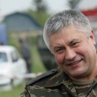 Vladimir Kolokoltsev: service in Afghanistan, the fight against organized crime groups and other facts from the biography of the head of the Ministry of Internal Affairs
 What is the difference between shallots and onions? Is it possible to replace them with each other? Differences from onions
 All women are like women, but I am a million dollar fool. And the main question is: what kind of woman do you need to be for this?
 Elsa Scarlett Titania. Rearmament of Elsa. Secrets of Erza Scarlett's Heart
 Anime character Elsa Alaya: biography, armor and interesting facts
 The most popular anime heroine