Angiofibroma in the nose from what it appears. Juvenile angiofibroma. Angiofibroma of the skin: manifestations of pathology and the degree of its danger
This type of tumor may not signal its presence for some time. Since the sooner a problem is detected, the easier it is to eliminate it, if incomprehensible nodules on the skin or discomfort in the nasopharynx form, you should contact specialists for diagnosis and, if necessary, treatment.
concept
If the formation is of a benign nature and it consists of connective and vascular tissue fibers, then experts define this phenomenon as angiofibroma.
A tumor of this type, among other pathologies, is considered rare. Usually develops with the same frequency in men and women.
More often, pathology is detected after forty years. In boys at the time of sexual development, juvenile angiofibroma may also occur. In these cases, education sometimes disappears when the youth reaches a period of maturity.
The tumor is located:
in rare cases on the face, in the respiratory tract on the mucous membranes, on the skin surface, more often in the area of \u200b\u200bthe extremities, in the kidney.
A tumor that forms in the nasopharynx often chooses the following dislocation:
pharyngeal fascia, sphenoid bone, ethmoid bone.
Causes of the disease
Angiofibroma on the skin occurs mainly in patients of mature age. Experts believe that the appearance of the described pathology on the skin occurs because the dermis undergoes photoaging.
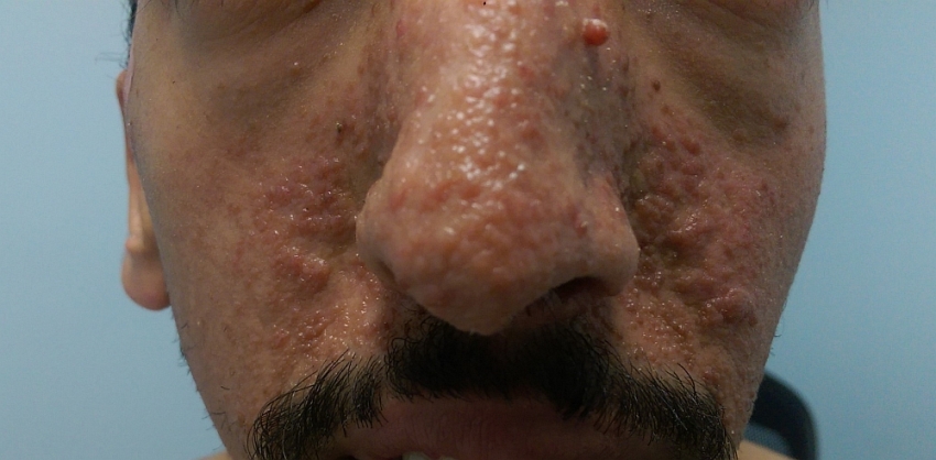
The photo shows multiple angiofibromas on the face of a young man
A tumor in the nasopharyngeal region is observed in young men when they have puberty.
It is believed that this process affects the mutation of cells in the nasopharyngeal zone. And the basis for the onset of the disease are the remains of embryonic tissue that have been preserved in this area in an undeveloped state.
Clinical picture
With the appearance of education in the nasopharynx, the picture of the disease can undergo four stages:
At the first stage, the presence of a localized tumor is observed. The second stage - the pathology begins to grow in the area of the sinuses. The bones of the nose may show signs of curvature. The third stage is characterized by the fact that the tumor process captures the region of the orbit and approaches the brain. There is a growth of education in the brain.
Symptoms of angiofibroma of the skin
Externally, the formation is a convex single node. Surface color:
brown, pale pink, pale yellow.
The top cover of a tumor looks transparent. Capillaries are visible under the skin. To the touch, the formation of a dense consistency, but at the same time retains elasticity.
Pathology is not of particular concern. A sign of angiofibroma is a slight itching in the area of education.
Signs of angiofibroma of the nasopharynx
When an angiofibroma appears in the nasopharynx, depending on the direction of its spread, one or more of the following symptoms may appear:
the eyeball undergoes a shift, this situation can affect visual acuity; nasal congestion of a chronic nature is observed, the face becomes asymmetric, facial tissues look edematous, breathing through the nose is difficult, there are signs of insufficient blood supply to the brain, headaches are often disturbed, the sense of smell is weakened, near the tumor the bone tissue undergoes deformation, nasal sound, nosebleeds appear, hearing loss is observed , nerve endings fall into a situation of compression by a growing tumor.
Diagnostics
To investigate a neoplasm on the skin that is similar to angiofibroma, the following actions are performed:
Visual examination of the tumor. For a detailed study of the node, a dermatoscope is used. The device gives an increase in the object hundreds of times. To determine the nature of the neoplasm in relation to malignancy, material is taken for histological examination. A general blood test will show the state of the body and determine if there is anemia.
In the case of dislocation of education in the nasopharynx, the following studies are also carried out:
Magnetic resonance imaging allows you to study the tumor to the smallest detail. Computed tomography can also give a lot of information about the formation: what is the spread of the tumor, determine the exact boundaries of the neoplasm, clarify the localization of the pathology. Radiography is carried out in order to determine what space the pathology occupies and its size. The method is not accurate and perfect. Sometimes it turns out only to confirm the presence of a tumor, it is better to obtain detailed information in another way. Rhinoscopy is performed anterior and posterior - it makes it possible to see the surface of the tumor and its color. Also, when touched with a probe, angiofibroma begins to bleed, which is important in determining the diagnosis. Nasal endoscopy allows you to perform a detailed examination of the condition of the nasopharynx and identify existing problems. It is carried out with the use of anesthesia.
Neoplasm treatment
An effective method of solving the problem when an angiofibroma is detected is its removal with a laser. The method of excision of the neoplasm is also used. Removal of the tumor is carried out to healthy tissues. In the area of the nasopharynx, access to the neoplasm is difficult due to the physiological features of the location of the pathology. In addition, the nasopharynx area has a large number of blood vessels arranged in a grid. The following types of operations are used: The intranasal endoscopic method is the most modern and atraumatic. From traditional methods: if the pathology has developed no higher than the second stage - lateral incision, if the tumor spreads more than the second stage - infratemporal incision.
To avoid bleeding during surgery, ligation of the carotid artery is often performed before surgery. If, nevertheless, blood loss has occurred, it is possible to replenish the volume of blood in the body by introducing donor material.
Surgery has a postoperative period. Appointments are being made:
often after surgery, radiation therapy is performed to remove the risk of infection, antibiotic therapy is used, and measures are taken to increase blood clotting. Radiotherapy is the irradiation of pathology with the help of special equipment. The method is used with caution. Experts recommend irradiating only the area with pathological cells and applying a precisely adjusted dose of radiation. This approach is called the stereotaxic technique. Hormone therapy uses testosterone to treat pathology. Studies have shown that this method leads to a cessation of tumor growth and a decrease in the size of the formation by almost half.
Complications after removal
When the pathology has sprouting into neighboring tissues, it is difficult to excise it, which can lead to some consequences:
tissues surrounding the tumor may be injured; possibly bleeding.
Radiation therapy is also fraught with consequences, it can happen:
skin atrophy, loss of appetite, decrease in blood erythrocytes and leukocytes, manifestation of dermatitis and skin edema.
Forecast
After removal of the tumor, its renewal is rare. It also rarely happens that a relapse leads to the fact that the resulting tumor acquires signs of a malignant formation.
Usually surgery results in a cure for the patient. Such treatment necessarily has a complex character: surgery and irradiation of the pathology site.
A neoplasm of connective tissues and blood vessels is considered a rather rare disease. In oncology practice, very often angiofibroma considered in combination with dermatofibroma. The localization of this benign tumor is the skin and the nasopharynx.
Causes and epidemiology of the disease
Angiofibroma of the nasopharynx was first described by Hippocrates in the 5th century BC. e. But the disease began to be called this term after 1940. The mutation of the cells of the nasopharyngeal space is mainly diagnosed in male patients aged 7-14 years, which is obviously associated with puberty.
Angiofibroma of the skin develops with the same frequency in both men and women. This skin lesion is the result of photoaging of the dermis. That is why older people are considered the most susceptible category.
Angiofibroma of the larynx: clinical picture
Symptoms of the disease include the following signs:
Chronic nasal congestion, which manifests itself in 80-90% of cancer patients in the early stages of the malignant process. Periodic nosebleeds. Blood discharge, as a rule, is unilateral and intense. This symptom is observed in 45% of clinical cases. Frequent headaches, which are provoked by constant congestion of the paranasal sinuses. Swelling of facial tissues. Juvenile angiofibroma with a significant spread, it can provoke a violation of auditory and visual functions.
Symptoms of angiofibroma of the skin
The pathological focus has the form of a dense node, the size of which does not exceed 3 mm in diameter. The color of the tumor can vary from light to dark brown. Such compaction of the epidermis in most cases does not cause subjective complaints in the patient and can be in a stable state for a long time.
Diagnosis of the disease
Atypical dermal tissue growth is diagnosed based on visual examination, which may be improved by dermatoscopy. The final diagnosis is established by the results of histological analysis. To conduct a biopsy, a small area of the oncological focus is removed from the patient and a laboratory analysis of the biopsy is performed.
Juvenile angiofibroma detected using the following methods:
Instrumental examination of the nasal cavity and pharynx. Computed and magnetic resonance imaging. Layered radiological scanning of an atypical area of the body identifies the boundaries, localization and spread of the neoplasm. Biopsy. A cytological biopsy test is necessary to clarify the diagnosis and type of tumor.
Differential Diagnosis
The cutaneous form of the pathology has a very similar clinical picture with dermatofibroma and melanoma.
Angiofibroma in a child differentiates with polyposis overgrowth, sinusitis and nasopharyngeal cancer.
Angiofibroma of the nasopharynx: treatment
Therapy of angifibroma lesions of the nasopharyngeal space is carried out by the following methods:
hormone therapy
Drug treatment includes the use of testosterone, which blocks tumor growth and causes a 44% reduction in tumor growth.
Radiotherapy
Some cancer centers report a positive result of radiological exposure in 80% of cancer patients. The use of radiation therapy has some limitations due to the high frequency of radiological complications. In this regard, oncologists recommend using a stereotaxic technique, which consists in a highly accurate and dosed behavior of radiation to the affected area of the body.
Surgery
Removal of angiofibroma is often complicated by the presence of a dense network of blood vessels. Surgical access to the pathological focus of the nasopharynx is carried out depending on the characteristics of the localization of oncology. For example, lateral nasal incision is indicated for stage 1 and stage 2 tumors; the infratemporal path is used for significant expansion of angiofibroma. Recently, intranasal endoscopic surgery has been widely used, with the help of which the surgeon excised a neoplasm with minimal trauma to nearby healthy tissues.
Consequences and complications after removal
Despite the key importance of surgical removal of the tumor, radical excision is contraindicated in 10% of clinical cases due to the growth of a benign neoplasm into the bone structures of the skull base. The main complications of such treatment are associated with recurrence of oncology (30% frequency), surgical bleeding and traumatic damage to adjacent tissues.
The effects of radiation therapy are as follows:
The development of radiological inflammation of the mucous membranes, in particular, stomatitis of the oral cavity. Decrease in the concentration of leukocytes and erythrocytes in the blood. Skin complications in the form of dermatitis, itching and swelling. Systemic manifestations of radiation intoxication (insomnia, loss of appetite).
Long-term effects of radiotherapy include atrophy of the skin, asymmetry of the facial skeleton, progressive osteoporosis and the formation of a secondary cancerous tumor.
life forecast
The prognosis of the disease is usually favorable. Timely surgical operation in combination with radiation therapy leads to a complete cure for a cancer patient.
In rare cases, a negative result of anticancer treatment is observed in the form of recurrence or malignancy of the neoplasm. Statistically, angiofibroma after resection undergoes cancerous transformation in the second or third year of the rehabilitation period. For timely diagnosis of therapeutic complications, patients are recommended to undergo annual examinations by an otolaryngologist.
Inna Bereznikova
Reading time: 5 minutes
A A
- a benign tumor. Most often, it looks like a skin formation of a pale pink or flesh color. Does not cause any inconvenience to a person, except perhaps cosmetic. It can be in a child from birth or it can be formed during life. Such a disease can be located anywhere, completely independent of gender or age. However, inflammation often occurs within organs. There are many types of fibroids, we will look at some of them.
Dermatofibroma or in another way is a neoplasm of connective tissue and skin, which sometimes looks like a wart. Dermatofibroma most often has a round shape, the main part of the inflammation is in the skin. Usually, dermatofibroma is a single formation, but there have been cases when several neoplasms appeared at once. Most often found on the arms, legs, neck, face and other parts of the body. Dermatofibroma has been seen most in middle-aged women.
Doctors associate the causes of this inflammation with several factors:
- polluted environment;
- heredity;
- infection;
- permanent skin injury.
Dermatofibroma can be dangerous, especially when it occurs in children. This is explained by the fact that with constant irritation, painful sensations can occur. The following symptoms can be considered signs of inflammation such as dermatofibroma.
- bleeding due to mechanical damage;
- if pressed, the dermatofibroma will bend inward;
- hypersensitivity, painful sensations when touched, as well as itching are not excluded.
Dermatofibroma is treated in only one way - removal, either laser or surgical.
It is important to know! Any neoplasm that has been removed, including dermatofibroma, needs a morphological analysis of tissues.
Angiofibroma is a benign neoplasm that consists of fibrous and vascular tissue. The diameter of angiofibroma is from three millimeters to several centimeters. The surface of the tumor itself is transparent, capillaries are visible, they are especially well visible during dermoscopy. The disease develops slowly, as a rule, angiofibroma takes on a progressive character after a person reaches the age of forty to fifty years. Angiofibroma is a rare skin disease.

Angiofibromas in tuberous sclerosis
As a rule, angiofibroma is formed on the skin of the extremities, but there is a case when the tumor appeared on the mucous membranes of the respiratory tract. There is no tendency to malignancy in this pathology. Angiofibroma in terms of its diagnosis is no different from other types of fibromas. To make an accurate diagnosis, it is necessary to conduct a histological examination, and only after an affirmative result, it is necessary to draw up a course of treatment.
Desmoid fibroma
Desmoid fibroma is an extremely rare disease, usually affecting the walls of the abdominal cavity. For a long time there was only one method of treatment - surgical, but with the development of medicine, combined methods appeared that effectively affected the tumor without surgical intervention.
Odontogenic fibroma
Odontogenic fibroma is a rare tumor located in the jaw area. Its main difference from other similar diseases is the presence of a cyst-like cavity with multiple dense inclusions.
Fibroma of the kidney
Kidney fibroma is one of, but it still cannot be started. Not always, other reasons are possible. If you notice symptoms of this pathology in yourself, we strongly recommend that you contact a medical specialist. Many believe that this disease can be cured only through traditional medicine, conspiracies and other non-medical methods. This opinion is wrong. Of course, there were cases when a person was able to recover using only traditional medicine, but such cases are extremely rare. With modern medicine, to refuse her help is at least stupid.
Chondromyxoid fibroma
Chondromyxoid fibroma is an extremely rare cartilage-forming inflammation. As a rule, it most often occurs in the metadiaphyses and metaphyses. You can also progress in other bones of the human body. The prognosis for a disease such as chondromyxoid fibroma is favorable, but there is one drawback. Chondromyxoid fibroma is prone to recurrence, and cancer may sometimes develop. Relapses occur in ten to fifteen percent, usually in the first two years after surgery.  Chondromyxoid fibroma is treated only with a surgical method. Marginal resection is usually used, which guarantees a minimum number of recurrences. If not on time, chondromyxoid fibroma can become malignant. Therefore, we strongly recommend that you properly respond to the manifestation of symptoms.
Fibroma of the bone
Bone fibroma is one of the most common tumor diseases. Usually, . It is noteworthy that after the operation for inflammation, neither a scar nor a seam remains. Therefore, the treatment of this pathology passes, most often, without complications.
Non-ossifying fibroma
Non-ossifying fibroma and metaphyseal defect are one of the most common skeletal diseases in childhood. These terms can be considered as synonyms, since they have almost no difference in their lexical meaning and mean the same thing. Do not be afraid of doctors or do not follow their recommendations and advice at all. In our time, medicine has reached such a level that it is able to remove any type of fibroma without consequences, whether it is a non-steggy fibroma or an unossified one, or any other. Many do not even know what a puncture is, but our medicine has been using it for a long time! Inflammation of the prostate or pancreas, soft tissues, in the pelvis, auricles, tibia, tendon, on the nose, near the eyes, in the abdomen or some other. Our medicine can handle everything! 

The most important thing to remember is to always listen to your healthcare professional.
If he says that these pills must be taken for 12 weeks, then this is how it should be done. Many decide for themselves when they can stop taking pills - this is wrong. If your symptoms are gone, it does not mean that you are completely healthy. It is necessary to adhere to a healthy diet, as well as a postoperative course of prevention. Particular attention should be paid to prevention in case of pregnancy. If you manage to move from diagnosing a disease to undergoing a course of treatment in time, you have nothing to worry about!
|
What is myoma? Its types and methods of treatment |
Types of benign and malignant melanoma |
Angiofibroma is a rare disease, it is formed from blood vessels and connective tissue. In oncology, angiofibroma is often considered in combination with dermatofibroma.
More about angiofibroma
The locations of this tumor are most often the nasopharynx and skin, although there are other localization sites - tendons, mammary glands, trunk, neck, face, ovaries, uterus, lungs, vocal cords.
Note! Usually, angiofibroma of the skin is diagnosed in males after 40 years, but it can also form in women. This type of neoplasm is associated with photoaging of the dermis, therefore, people of the older generation are included in the risk group.
In children during puberty, an angiophyroma of the juvenile type (youthful) can be diagnosed, which can go away on its own with age.
Remember! Usually, juvenile angiofibroma is diagnosed in the nasopharyngeal zone - this is the so-called mutation of the nasopharyngeal space.
Leading clinics in Israel
Causes
The exact cause of angiofibromas has not been established, but it is generally accepted that abnormal development of the embryo plays an important role here.
In general, there are several theories of the occurrence of this disease:
- genetic. Refers to the most common, since the vast majority of patients diagnosed with angiofibroma find chromosomal abnormalities;
- hormonal. Frequent diagnosis of one of the types of angiofibroma in adolescents during puberty, gives reason to assume the cause of the formation of angiofibroma - hormonal imbalance;
- age. The risk of getting this disease increases with age, given the natural aging process of the body.
In addition to the above theories, there are assumptions regarding the effects of some other factors:

One of the main factors provoking the disease is considered to be prolonged exposure to sunlight (photoaging), which causes mutations in cells that are located in the deep layers of the skin. In the process of mutation, they begin to intensively divide and form a focus with pathologically developed blood vessels.
Sometimes there are multiple angiofibromas (angiofibromatosis), which can be the result of hereditary diseases - neurofibromatosis and tuberous sclerosis.
Symptoms of the disease
Symptoms of the disease include manifestations that depend on the location of the angiofibroma:
- in most cases (80-90%) in the early stages of the disease, chronic nasal congestion occurs (with angiofibroma in the nasopharynx);
- almost half of the cases (45%) may experience nosebleeds. More often they are unilateral;
- due to congestion of the paranasal sinuses, frequent headaches may occur;
- if juvenile angiofibroma has a significant distribution, then it can provoke violations of visual and auditory functions;
- when localized in the face area, its swelling may occur, facial asymmetry can be observed.
In some cases, symptoms are not observed (with kidney damage).
Outwardly, this neoplasm may look like a single node (up to 3 mm in size), rising above the skin. Its color can be from almost indistinguishable from skin color to dark brown, and the upper layer of the formation is transparent and makes it possible to see the capillary pattern from small vessels. The knot itself is slightly tighter than the surrounding skin, but is quite elastic. Although the knot itself is painless, it is accompanied by mild itching. The tumor develops slowly and does not cause changes in surrounding tissues.
Tumor types
Depending on the microscopic structure, cutaneous angiofibromas can be divided into:
- hypercellular. They consist of a large number of fibroblasts - immature connective tissue cells;
- typical (angiofibrolipoma). These types of tumors are composed of foam cells that contain lipids;
- pigmented. Such a tumor has many melanin pigments in its composition, and is often mistaken for a mole;
- pleomorphic. This form contains nuclei of different shapes and sizes, and this sometimes leads to an incorrect diagnosis - a malignant skin tumor is determined - a sarcoma;
- granular (angiogranuloma). The internal environment of such cells has granules, because of which they can be confused with malignant ones.
According to the place of localization, this disease can be divided into:
Depending on the clinical and anatomical features, angiofibroma can be distinguished:
- basal-common;
- intracranially widespread.
If we consider in more detail angiofibroma of the nasopharynx, then There are several stages of its development:
- stage 1 - the tumor does not go beyond the boundaries of the nasopharynx;
- 2 - pathological tissues extend to the pterygopalatine fossa, into some sinuses (maxillary, sphenoid, ethmoid);
- Stage 3 can occur in two versions: 1 - the pathology begins to spread to the region of the orbit and to the infratemporal region, 2 - the hard shell of the brain is involved in the process;
- Stage 4 can also occur in two variants: the first variant is characterized by damage to the dura mater, but without being drawn into the pathological process of the pituitary gland, optic chiasm or cavernous sinus, the second variant is characterized by the spread of the tumor to all of the above areas.
Diagnosis of the disease
 Pathological proliferation of skin tissues can be diagnosed on the basis of an external examination, which is carried out in conjunction with. The final diagnosis is based on the results of histology. For a biopsy, a small area of the pathological focus is removed and a laboratory analysis of the biopsy is done.
In addition to these diagnostic methods, palpation of suspicious areas of the body, MRI (it identifies the boundaries of the formation, localization and spread of the tumor), anterior and posterior rhinoscopy, radiography, ultrasonography, CT, fibroscopy, angiography are carried out. Various laboratory tests are also carried out: blood, blood biochemistry, hormonal tests.
Important! If an angiofibroma is suspected, cancer must first be ruled out. They also perform differential diagnosis with diseases, such as: hemangiomas, lipomas, nevi, melanoma,. In children, angiofibroma is differentiated from polyposis overgrowth, nasopharyngeal cancer and sinusitis.
Angiofibroma treatment
Therapy of angiofibroma of the nasopharynx can be carried out by the following methods:
- hormone therapy. Hormone treatment includes the use of testosterone, which blocks tumor growth and causes tumor shrinkage by 44%;
- radiotherapy. This type of treatment has some limitations due to the high percentage of radiological complications. Doctors often advise using a stereotaxic technique, which uses highly precise and dosed use of radiation;
- surgical intervention. Due to the presence of a dense network of blood vessels, removal surgery is not always possible. In recent years, intranasal endoscopic surgery has been more frequently used, when the surgeon performs excision of the tumor with minimal damage to nearby normal tissues.
After removal, the tumor almost never recurs.
*Only subject to obtaining data on the patient's disease, a clinic representative will be able to calculate an accurate estimate for treatment.
Consequences after surgery
Sometimes, after surgery, relapses of the disease, surgical bleeding and damage to neighboring tissues are possible.
After radiotherapy, the following complications may occur:

Disease prognosis and prevention
The prognosis of the disease is favorable. A timely surgical operation in combination with radiotherapy gives a high chance of curing the patient. In exceptional cases, there may be a relapse of the disease or malignancy of the tumor. Statistics say that after an angiofibroma is removed, it can undergo cancerous degeneration in the 2-3rd year of the rehabilitation period. For timely diagnosis of the disease, it is necessary to undergo annual preventive examinations by an otolaryngologist.
- maintaining a healthy lifestyle;
- proper nutrition;
- giving up bad habits;
- avoiding nervous and physical overstrain.
skin fibroma- this is a benign neoplasm of the skin, in the form of a nodule with a diameter of 0.2-1.5 cm, usually the color of normal skin, densely elastic consistency - hard fibroma or in the form of a soft "skin bag" on the leg - soft fibroma. The tumor consists of connective tissue, in its structure it can have muscle, vascular, adipose tissue. Fibroma, angiofibroma are derivatives of this tumor.
Angiofibroma-
it is a benign skin lesion, usually in the form of a pronounced densely or softly elastic nodule with a diameter of 0.3-1.5 cm, skin color or yellowish-brown, consists of connective tissue with vessels on the surface. The vascular network on the surface, sometimes, gives a pinkish appearance to angiofibroma. Vessels of angiofibroma can have a different look - commas,  convoluted lines, dots, which is clearly visible during dermatoscopy.
Angiofibroma- a common skin tumor and occurs equally often in both men and women, usually after 40-45 years. The cause of angiofibroma is photoaging of the skin or a genetic predisposition. Angiofibroma is located on the skin of the extremities, back, face, neck.
Angiofibroma rarely degenerates into a malignant tumor. However, having vessels of different structure on the surface in its structure, it can hide under the mask of angiofibroma. Therefore, if you notice this kind of neoplasm in yourself or your loved one, immediately contact an experienced one. So  angiofibroma must be differentiated from dermatofibroma, fibroma, pigmented nevus, melanoma, angioma.
Appears fibroma skin at any age, but more often after 30 years, both in men and women. Located T verdaya fibroma more often on the face, neck, back, chest, limbs. Soft fibroma is located in the inguinal, axillary folds, under the breasts in women, on the neck.
Fibroma of the skin must be distinguished from other skin neoplasms such as angiofibroma, papilloma, dermatofibroma, basalioma. The tumor rarely degenerates into a malignant one.
Laser removal of skin fibroids is the most effective, safe, aesthetic method of treatment .


The disease "angiofibroma of the skin" is a benign tumor consisting of connective tissue fibers and blood vessels, located on the skin of the extremities, cheeks, in the nose, in the nasopharynx, and also in the kidneys.
The tumor usually develops in men over 40 years of age. Less commonly, it occurs in boys during puberty, and disappears with age. This form is called juvenile or juvenile angiofibroma.
Causes
The exact cause of the appearance of dermal angiofibromas has not been established, but it is believed that the action of direct sunlight plays an important role in this, that is. Solar radiation causes mutations in cells located in the deep skin layers. As a result, they begin to intensively divide and form a pathological focus, supplied with abnormally developed blood vessels.
Multiple cutaneous angiofibromas can be a sign of hereditary diseases such as tuberous sclerosis and neurofibromatosis.
Unlike malignant tumors, angiofibroma does not penetrate deeper than the lower dermal layers, its cells do not enter the vessels and lymphatic capillaries and do not form metastases. The course of the disease is benign. It is not contagious and does not threaten the life of the patient.
Symptoms
External signs of the disease do not always make it possible to accurately diagnose.
- Angiofibroma of the skin looks like a single small node protruding above the surface of the epidermis (from 1 mm to 1 cm, up to a maximum of 3 cm).
- It can have a different color - from light beige to brown or pink.
- The upper layer of the epithelium of the formation looks thin and translucent, through which the capillary pattern of small blood vessels is clearly visible.
- The consistency of the knot is denser than the surrounding skin, but at the same time it retains elasticity.
- Angiofibroma is painless, but may be accompanied by mild itching.
- The tumor grows very slowly, without causing changes in the surrounding tissues.
Diagnostics
If an angiofibroma of the skin is suspected, the doctor first of all seeks to exclude. It uses the following diagnostic methods:
- inspection, palpation;
- - examination under a special microscope, giving an increase of hundreds of times;
- examination of the formation in ultraviolet rays to identify accumulations of skin pigment in it;
- biopsy of the material and its histological examination;
- complete blood count - to detect anemia and other pathological changes characteristic of cancer.
Types of pathological formations
Depending on the microscopic structure, the following types of cutaneous angiofibromas are distinguished:
- hypercellular: consists of a large number of immature connective tissue cells - fibroblasts;
- a typical tumor with foam cells containing lipids;
- pigmented: contains a lot of skin pigment melanin, can be mistaken for a mole;
- pleomorphic: cells have nuclei of different sizes and shapes, which can lead to an erroneous diagnosis of a malignant skin tumor - sarcoma;
- granular: the internal environment of the cells has granules, as a result of which they can also be confused with malignant.
Differential diagnosis is carried out with other benign and malignant neoplasms of the skin:
- birthmark;
Treatment
Although such a neoplasm does not metastasize, it can be damaged by rubbing against clothing, become inflamed, lead to bleeding, or cause aesthetic discomfort. Therefore, the treatment of angiofibroma of the skin is to remove it.
If the focus is located in a place that is not injured, or it does not lead to defects in appearance, then after diagnosis, you can limit yourself to periodic observation by a dermatologist, without removing it.
Angiofibroma can be removed in different ways:
- Surgical excision with a scalpel
The procedure is performed on an outpatient basis under local anesthesia. The surgeon removes the tumor node and applies several small stitches, which later dissolve. The advantage of the method is the complete excision of the formation and the possibility of subsequent histological analysis of the obtained tissues. The disadvantages include the formation of a small skin scar, the possibility of bleeding or infection.
- laser removal
With the help of a laser beam, the doctor “evaporates” the tissues of the node in layers, reaching healthy tissues. The treatment does not cause blood loss, is not accompanied by infection, and does not leave a scar after it. Laser tumor removal is performed under local anesthesia and lasts no more than 10-15 minutes. This is the most modern way to treat angiofibromas.
- Radio wave treatment
Using a beam of radio waves, the doctor completely removes the pathological formation, while there is no bleeding, and the scar after such a procedure is absent or almost invisible. However, radio wave treatment can lead to local skin atrophy or development.
To improve the regeneration of skin tissues after any type of angiofibroma removal, it is necessary to exclude friction of the operated area, change the bandage in time, and follow the doctor's recommendations to prevent inflammation.
You can take antibiotics, use wound healing ointments only on the advice of a doctor. Usually this is not advised, since the area of intervention is very small, and the wound heals quickly under a bandage.
After removal, the tumor almost never recurs. Recurrence is usually a sign of previously unrecognized skin cancer. If multiple angiofibromas are associated with a genetic disease, then the prognosis is determined by the presence of the underlying pathology.

Contraindications for removal
There are no absolute contraindications to the removal of angiofibroma, if it is really necessary. However, there are relative contraindications in which it is better to transfer the procedure until you feel better:
- acute infectious diseases, febrile conditions;
- decompensated course of diabetes mellitus with high blood sugar levels;
- immunodeficiency states, long-term use of glucocorticoids, polychemotherapy for malignant tumors;
- dermatitis or skin damage in the area of intervention;
- pregnancy, menstrual bleeding;
- when using surgical excision - a tendency to bleeding or the formation of rough skin scars.
 Ancient and very strange sports Pistol duel
 The oldest sport in the world
 Vladimir Kolokoltsev: service in Afghanistan, the fight against organized crime groups and other facts of the biography of the head of the Ministry of Internal Affairs
 Foster Parent School
 Super bright LEDs. Types and device. Work and application. Selection, application and characteristics of LEDs: useful information and professional recommendations LED super bright powerful white
 All women are like women, and I am a fool for a million And the main question is: what kind of woman do you need to be for this
 Elsa scarlett titania. Elsa rearmament. Erza Scarlett's Heart Secrets